DIAGNOSIS
Because there are currently more than 1,200 disorders and chromosomal abnormalities that can be diagnosed by looking for a specific change in a patient's DNA (GeneTests, 2008), the art of medical genetics is an important part of patient care. Medical geneticists must therefore have a clear understanding of the different types of diagnostic tests available to patients, including biochemical testing, karyotyping, FISH, preimplantation genetic diagnosis, and newborn genetic screening.
Biochemical Testing
Sometimes, a pediatrician will suspect that a child has a genetic disorder based on the child's symptoms or on the presence of dysmorphic features. For example, if a child has coarse facial features and developmental delays, a pediatrician may have reason to believe that the child has a form of mucopolysaccharidosis. Mucopolysaccharidosis is a family of diseases caused by an enzyme deficiency that leads to the accumulation of glycosaminoglycans (GAGs) within the lysosomes of cells. In one particular variant of this disease known as mucopolysaccharidosis I (MPS I), a deficiency of the enzyme alpha-L-iduronidase causes a build up of GAGs in tissues and organs, which in turn leads to a host of signs including skeletal deformities, coarse facial features, enlarged liver and spleen, and mental deficiencies. Because of the progressive nature of MPS I, a child might not exhibit noticeable symptoms until one to three years of age or even later, depending on severity.
There are a number of reasons that a pediatrician might refer a child to see a geneticist. Geneticists can confirm or rule out a physician's diagnosis based on the findings of a physical exam and various tests. In the case of a child with suspected MPS, if the enzymatic deficiency associated with the disorder is confirmed via testing, DNA analysis may also be performed to determine the exact genetic mutation causing the disorder. Because MPS I is inherited in an autosomal recessive fashion, identification of the mutation can allow the family to undergo carrier screening, as well as prenatal or preimplantation diagnosis in any future children.
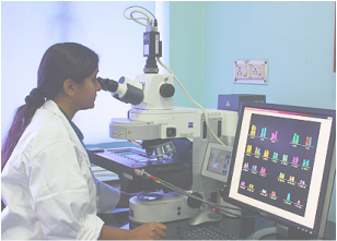
Karyotyping and FISH
In other cases, a physician might suspect a chromosomal abnormality prior to birth. For example, an obstetrician may suspect that a fetus has Down syndrome based on maternal blood testing or findings on an ultrasound examination. Individuals with Down syndrome have characteristic facial features and lower than average cognitive abilities, and they are also at higher risk for heart defects and other medical problems. The most common cause of Down syndrome is trisomy 21 (i.e., the presence of three copies of chromosome 21), a condition that results from a meiotic nondisjunction event, usually in the mother. The risk of this type of trisomy 21 increases with maternal age.
One way to test for Down syndrome is to karyotype fetal DNA; this involves obtaining fetal cells via amniocentesis, then culturing the cells and staining the chromosomes so that they can be visualized under a microscope. A second testing method is fluorescence in situ hybridization (FISH). In this technique, labeled DNA probes complementary to regions of the chromosome in question are allowed to hybridize to a preparation of the test sample's chromosomes. A hybridization signal verifies the presence of that chromosomal material in the test sample, while the absence of a signal indicates the absence of the material. Diagnostic FISH for Down syndrome would involve the use of labeled probes with sequences complementary to regions spanning chromosome 21. The presence of three labeled chromosomes would therefore be diagnostic of Down syndrome. Both of these techniques have their benefits and limitations.
Preimplantation Genetic Diagnosis (PGD)
Genetic testing can also be conducted at the embryonic stage, before implantation. For instance, parents who are both carriers of an autosomal recessive disorder, such as cystic fibrosis (CF), have a 25% chance with each pregnancy of having a child with CF. Such parents may want to take advantage of modern technology that enables screening for genetic diseases during the earliest stages of embryonic development. One example of this type of technology is preimplantation genetic diagnosis (PGD), a procedure in which individuals undergoing in vitro fertilization have their developing embryos tested for known genetic abnormalities prior to implantation in the mother's uterus (Handyside et al., 1992). In PGD for cystic fibrosis, for example, one or two cells would be removed from the early embryos in vitro and tested for the specific CF mutations carried by the parents. Only embryos determined to be absent of these mutations would then be transferred to the mother's uterus, where they would subsequently undergo development (Figure 1).
Newborn Genetic Screening
Finally, as part of a public health initiative, babies may be diagnosed with genetic disorders without any suspicion at all. This is called newborn screening. Here, a small blood sample is collected from newborn infants within 24 hours of birth and tested for a panel of disorders. The American College of Medical Genetics (ACMG) recommends a core screening panel for 29 conditions, but the actual number of conditions that most facilities test for varies by state and country, ranging from as few as three to more than 40 (MedlinePlus, 2008). Receiving a genetic diagnosis from a newborn screen like this can save an infant's life, because the disorders included in the panel are those in which early diagnosis and treatment are imperative for a good outcome. One such disorder is phenylketonuria (PKU), a condition in which affected individuals are unable to properly metabolize the amino acid phenylalanine. Over time, this substance can therefore accumulate in the person's body, leading to a host of problems, including possible mental retardation. Thankfully, PKU can be treated through adherence to a strict diet that is low in phenylalanine, especially early in life. PKU is one of the most prominent success stories showing that early diagnosis and tailored treatment of genetic disorders can improve a person's quality of life.
Biochemical Testing
Sometimes, a pediatrician will suspect that a child has a genetic disorder based on the child's symptoms or on the presence of dysmorphic features. For example, if a child has coarse facial features and developmental delays, a pediatrician may have reason to believe that the child has a form of mucopolysaccharidosis. Mucopolysaccharidosis is a family of diseases caused by an enzyme deficiency that leads to the accumulation of glycosaminoglycans (GAGs) within the lysosomes of cells. In one particular variant of this disease known as mucopolysaccharidosis I (MPS I), a deficiency of the enzyme alpha-L-iduronidase causes a build up of GAGs in tissues and organs, which in turn leads to a host of signs including skeletal deformities, coarse facial features, enlarged liver and spleen, and mental deficiencies. Because of the progressive nature of MPS I, a child might not exhibit noticeable symptoms until one to three years of age or even later, depending on severity.
There are a number of reasons that a pediatrician might refer a child to see a geneticist. Geneticists can confirm or rule out a physician's diagnosis based on the findings of a physical exam and various tests. In the case of a child with suspected MPS, if the enzymatic deficiency associated with the disorder is confirmed via testing, DNA analysis may also be performed to determine the exact genetic mutation causing the disorder. Because MPS I is inherited in an autosomal recessive fashion, identification of the mutation can allow the family to undergo carrier screening, as well as prenatal or preimplantation diagnosis in any future children.
Karyotyping and FISH
In other cases, a physician might suspect a chromosomal abnormality prior to birth. For example, an obstetrician may suspect that a fetus has Down syndrome based on maternal blood testing or findings on an ultrasound examination. Individuals with Down syndrome have characteristic facial features and lower than average cognitive abilities, and they are also at higher risk for heart defects and other medical problems. The most common cause of Down syndrome is trisomy 21 (i.e., the presence of three copies of chromosome 21), a condition that results from a meiotic nondisjunction event, usually in the mother. The risk of this type of trisomy 21 increases with maternal age.
One way to test for Down syndrome is to karyotype fetal DNA; this involves obtaining fetal cells via amniocentesis, then culturing the cells and staining the chromosomes so that they can be visualized under a microscope. A second testing method is fluorescence in situ hybridization (FISH). In this technique, labeled DNA probes complementary to regions of the chromosome in question are allowed to hybridize to a preparation of the test sample's chromosomes. A hybridization signal verifies the presence of that chromosomal material in the test sample, while the absence of a signal indicates the absence of the material. Diagnostic FISH for Down syndrome would involve the use of labeled probes with sequences complementary to regions spanning chromosome 21. The presence of three labeled chromosomes would therefore be diagnostic of Down syndrome. Both of these techniques have their benefits and limitations.
Preimplantation Genetic Diagnosis (PGD)
Genetic testing can also be conducted at the embryonic stage, before implantation. For instance, parents who are both carriers of an autosomal recessive disorder, such as cystic fibrosis (CF), have a 25% chance with each pregnancy of having a child with CF. Such parents may want to take advantage of modern technology that enables screening for genetic diseases during the earliest stages of embryonic development. One example of this type of technology is preimplantation genetic diagnosis (PGD), a procedure in which individuals undergoing in vitro fertilization have their developing embryos tested for known genetic abnormalities prior to implantation in the mother's uterus (Handyside et al., 1992). In PGD for cystic fibrosis, for example, one or two cells would be removed from the early embryos in vitro and tested for the specific CF mutations carried by the parents. Only embryos determined to be absent of these mutations would then be transferred to the mother's uterus, where they would subsequently undergo development (Figure 1).
Newborn Genetic Screening
Finally, as part of a public health initiative, babies may be diagnosed with genetic disorders without any suspicion at all. This is called newborn screening. Here, a small blood sample is collected from newborn infants within 24 hours of birth and tested for a panel of disorders. The American College of Medical Genetics (ACMG) recommends a core screening panel for 29 conditions, but the actual number of conditions that most facilities test for varies by state and country, ranging from as few as three to more than 40 (MedlinePlus, 2008). Receiving a genetic diagnosis from a newborn screen like this can save an infant's life, because the disorders included in the panel are those in which early diagnosis and treatment are imperative for a good outcome. One such disorder is phenylketonuria (PKU), a condition in which affected individuals are unable to properly metabolize the amino acid phenylalanine. Over time, this substance can therefore accumulate in the person's body, leading to a host of problems, including possible mental retardation. Thankfully, PKU can be treated through adherence to a strict diet that is low in phenylalanine, especially early in life. PKU is one of the most prominent success stories showing that early diagnosis and tailored treatment of genetic disorders can improve a person's quality of life.
